Scientific Program

Day 1 :

Keynote Forum

Diana Hodgins

European Technology for Business Holdings Ltd, UK

Keynote: Changing the care pathway for hip and knee replacement patients

Time : 10:00-10:40

Biography:

Diana obtained her degree in Mechanical Engineering and her PhD in solid state gyroscopes from the University of Hertfordshire (UH). Diana has 30 patents granted on solid state sensors and more recently on medical applications relating to the use of these sensors. In 2000 Diana was awarded an MBE for services to SMEs in the region and in 2002 she won the Women Inventor of the Year Award for Industry. In 2005 Diana was awarded an Honorary doctorate from the UH for services to innovation. In 1995 Diana and her husband established their own business, and in 2009 launched their sensor based gait monitoring product, GaitSmart. This system is now in use in variety of healthcare and sports applications. One of the sectors where it is applied is orthopaedics, with a particular focus on osteoarthritis and a number of papers have been published on this topic. Diana is currently a visiting professor at the UH and helped to establish a new MEng course in Biomedical Engineering and in 2014 was awarded Alumni of the year at the UH.

Abstract:

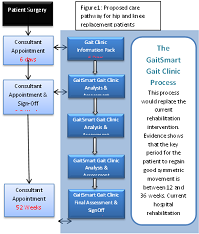
People with late stage hip or knee osteoarthritis may receive a joint replacement to relieve pain and improve mobility.  The operation is considered a success, with regard to patient reported outcomes.  However, studies using GaitSmart on hip and knee replacement patients one year post op show that less than 50% return to a normal gait and that these gait deficiencies can result in osteoarthritis in other joints, poor mobility and even falls if not corrected. People adapt their gait due to pain in the arthritic joint.  Once the joint pain is gone, the patient needs retraining to walk correctly and weakened muscles strengthened.  The current care pathway provides patients with physiotherapy for up to 6 weeks post op, guiding the patient to move the joint and become mobile.  However, as it takes 10 weeks for the process of muscle strengthening to start, patients are signed off before gait retraining can really start. Patients are unaware of how they have adapted their gait; for example, excessive medial-lateral movement of the thigh i.e swinging the leg around the body rather than under the body.  Therefore, the walking pattern remains the same, loading other joints incorrectly and poor use of their muscles. This paper proposes a new care pathway for patients, where gait kinematics are measured in the outpatient clinic using inertial sensors.  A report describing the gait deficiency and the severity, using simple traffic light coding, together with a personalised exercise programme is provided to the patient at the end of their 15 minute session.  Tests performed at 6, 12, 24 and 52 weeks post op monitor progress and allow the exercises to become more demanding as the patient improves. Evidence of the clinical efficacy of this approach will be presented.


Keynote Forum

Jike Lu

Beijing United Family Hospital, China

Keynote: The highly variable typologies of posterior malleolus fractures of the ankle

Time : 10:40-11:20

Biography:

Dr. Jike Lu is an Orthopaedic surgeon, Chairperson, Professor, Dept of Orthopaedics at Wenzhou Med University, BJU, Beijing China. He completed his doctorate at the University of New South Wales in Sydney. Before his doctoral studies in Australia, Dr. Lu was a qualified orthopedic spine surgeon and Professor of Orthopedic Surgery at Xinjiang Medical University. Dr. Lu accumulated ten years of extensive experience in orthopedic trauma, tumors, joint replacement, and spinal surgeries. He has served in numerous hospital institutions in Melbourne and Sydney. Dr. Lu’s primary clinical interest is orthopedic surgery for adults and children. In addition, Dr. Lu has also served as a visiting professor of orthopedic surgery at Wenzhou Medical University. Dr. Lu has over 30 years of clinical experience in the treatment and diagnosis of spinal degenerative disorders, cervical spondylosis, lumbar disc herniation, stenosis and spondylolisthesis, and spinal trauma and tumors. Dr. Lu specializes in orthopedic trauma, joint arthroplasty, and total knee joint and hip replacement. Dr. Jike Lu has published over 50 Research articles in Chinese in scientific and medical journals. Dr. Lu currently holds unconditional national medical board registration and is certified by the Australia Medical Council.

Abstract:

Objectives: Although historical studies frequently classify posterior malleolus fractures (PMFs) according to fragment size, our hypothesis is that PMFs have more complex patterns than is widely recognized. None of the studies so far have provided a comprehensive picture of the complexity of PMFs or guidelines for surgical decision making. The optimal management of PMFs is still unclear. This study aims to look at the typology of PMFs using a combination of initial injury radiographs, preoperative computed tomographic (CT) scans, and intra-operative image intensifier (II) screening, as well as fixation technique and a surgery fixation approach, in order to gain insight into PMFs.
Methods: Between 2013 and 2015, 56 consecutive patients, all with bi- or tri-malleolar fracture or dislocation of ankle joints, with one or more posterior fragments, who were treated at our institute, were identified. We retrospectively analysed the patients’ preoperative initial injury radiographs, CT scans, and II data to see the stability of the ankle joints in coronal and sagittal planes, and look at the typology in relation to fracture fixation technique and surgery fixation approach.
Conclusions: Bi- or tri-malleolar fractures of the ankle with associated posterior malleolar fractures appear to be highly variable. We identified certain types of PMFs which we can categorise. Ankle stability in the coronal and sagittal planes on initial injury radiographs, intra-operative II and preoperative CT scans are critical in order to identify different PMFs pat- terns. PMFs have highly variable typology regardless of fragment size and this must take into consideration when deciding treatment plans.

Keynote Forum

Takatomo Mine

National Hospital Organization Kanmon Medical Center, Japan

Keynote: A kinematic analysis in posterior-stabilized total knee arthroplasty during activities of daily living

Time : 11:40-12:20

Biography:

Dr. Takatomo MINE was born in Fukuoka, Japan. He got his bachelor degree from Yamaguchi University School of Medicine. He got his Ph.D. (Dr. of Medical Science) degree at Yamaguchi University School of Medicine. He is currently an Associate Professor in the Department of Orthopedic Surgery, Yamaguchi University. He is the Director of Orthopedic Surgery & Rheumatology at Kanmon Medical Center, Yamaguchi and also a member of various associations: Japanese Orthopedic Association(1985), The Central Japan Association of Orthopedic Surgery & Traumatology(1985), International Society of Arthroscopy, Knee Surgery and Orthopaedic Sports Medicine(2014), Japanese Society for Regenerative Medicine(2015), Japanese Society for Cartilage Metabolism(2017) and many other. He has published around 35 articles in various International Journals in the field of Orthopedics.

Abstract:

Background: Stair stepping motion and standing & sitting motion from a chair are important in daily living, similar to gait. It is important to understand in vivo kinematics of patient’s with total knee arthroplasty during stair-stepping and standing & sitting motion from chair. The purpose of this analysis was to estimate in vivo knee motion in stair stepping and standing & sitting motion from a chair, and determine if this unique knee prosthesis function as designed.
Methods: A total of 20 patients implanted with Bi-Surface PS were assessed in stair-stepping. 15 patients were assessed in standing & sitting from chair. The Bi-Surface PS knee is a posterior-cruciate substitute prosthesis with a unique ball-and-socket joint in the mid-posterior portion of the femoral and tibial components. Patients were examined during stair stepping and standing & sitting motion from a chair motion using a 2-dimensional to 3-dimensional registration technique.

Results: In stair-stepping, the kinematic pattern in step up was a medial pivot, in which the level of anteroposterior translation was very small. In step down, the kinematic pattern was neither a pivot shift nor a rollback. From minimum to maximum flexion, anterior femoral translation occurred slightly. In standing & sitting from a chair, from minimum to 30° knee flexion, anterior femoral translation occurred slightly. From 30° knee flexion to maximum flexion, the kinematic pattern was a medial pivot and rollback.

Conclusion: It became clear in this study that the joint’s stability during stair-stepping was affected by the design of the femorotibial joint rather than Post/Cam engagement or the Ball & Socket joint. In standing & sitting from a chair, the unique knee prosthesis functioned as designed.

Keynote Forum

Margo J Apostolos

University of Southern California, USA

Keynote: Trauma no drama: Care and treatment of dancers

Time : 11:40 AM

Biography:

Margo K. Apostolos, PhD. is the Co-Director and Co-Founder of the Cedars-Sinai/ USC Glorya Kaufman Dance Medicine Center and Associate Professor of Dance at the University of Southern California. Apostolos earned a PhD in Physical Education with a Philosophy minor a Stanford University, MA in Dance from Northwestern University, and BS in Physical Education with a Dance minor from Southern Illinois University-Carbondale. Her practiced based work in training dancers and athletes spans over forty years including numerous Olympians and professional athletes. In addition, Apostolos pioneered work in the development of Robot Choreography and was awarded a prestigious NASA/JPL Faculty Fellowship for work in space telerobotics. She works with both humans and robots.

Abstract:

Dance Medicine, similar to Sports Medicine, treats the specialized needs of dancers.  The art of dance parallels sport with the human body as the vehicle for performance.  While the aesthetic of dance differs from sport, dancers train differently than athletes and often periodization is not implemented in dance training. As the performances near, rehearsals often increase at full speed.  Overtraining becomes a common cause of dance injuries but the choreography makes dance ever so risky. The choreography of today places new demands on the dancers.  The inclusion of aerial maneuvers, acrobatic work, and elaborate set designs raise both the risk for injury and the nature of those injuries.  Aerial work and acrobatic maneuvers are included in both concert and commercial performance.  The dance environments have become spectacular from Las Vegas stages, film special effects, and the raked stages of opera houses. Similar to athletes, dancers are not at risk for traumatic injuries. At the Dance Medicine Center, we have seen traumatic injuries resulting from both choreography and the dance environment. The needs of the dancer are different than the athlete.  It is critical for the medical community to understand dancers especially in care, diagnosis, and treatment. This presentation will feature a case study of a traumatic dance injury documented from onset to recovery.  In addition, video links of current choreography will provide orthopedic surgeons an insight into the world of dance today. 

Biography:

Dr. Jeong is the new chairperson of World Taekwondo medical and anti-doping committee who introduced the use of point-of-care ultrasound to World Taekwondo for the first time. He has an extensive background in the use of point of care ultrasound in primary care, sports medicine, emergency & critical care medicine. He is certified in diagnostic musculoskeletal (RMSK) and abdomen(RDMS) sonography as well as EM POCUS (APCA). He has written three book chapters on this subject and has developed curriculum in this area for international Taekwondo team doctors, attending physicians, fellows, resident physicians, and medical students. He has been invited as an instructor for nationally well-known ultrasound workshop programs at UC Irvine Annual Ultrafest and National Center for Procedures Institute Ultrasound workshop for primary care physicians.

Abstract:

Statement of the Problem:  Physicians volunteering for sporting events may face with many acute traumas and musculoskeletal injuries at sporting events. At many times, on-site imaging modality such as x-ray, CT or MRI is not available which leads to unnecessary transferring athletes to other facility just for imaging studies due to uncertainty of diagnosis. Also, it is often difficult to obtain imaging studies for the injured due to availability or cost especially at international competition events.  Recently, much smaller and cheaper portable ultrasound machines with better image quality has been available so many clinicians has been utilizing portable ultrasound in their practice for point-of-care clinical decision making. However, there is very few research on using this modality at sporting events.
Methodology & Theoretical Orientation: Since 2017 Muju World Taekwondo Championships in June 2017, World Taekwondo (WT) medical and anti-doping committee has been adopted point-of-care ultrasound (POCUS) project using portable ultrasound machines for acutely injured athletes in Taekwondo competitions at WT promoted events.
Findings: Injury surveillance using portable ultrasound results revealed that there were more fracture and high-grade ligament tear cases in upper compared to previously published epidemiology studies on Taekwondo competition-related injuries. Also, there were some findings requiring urgent referral such as traumatic vitreous detachment, comminuted nasal fracture, pneumothorax, comminuted tibial shaft fracture and acute DVT of peroneal vein. The on-site POCUS in competition enabled venue clinicians to make more accurate diagnosis which resulted in the decease of the number of unnecessary hospital transfer by 35% and detect serious injuries in timely manner. 
Conclusion and Significance:  The use of Portable ultrasound is very useful and valuable in acute trauma setting at sporting events to make faster and more accurate diagnosis which may improve the on-site management of acute sports-related injuries.

  • Orthopedic Trauma | Fracture | Orthopedic Surgery | Rheumatoid arthritis | Arthroplasty
Location: London, UK
Speaker
Biography:

Dr. Francisco J. Sobrino is a Medical specialist in Traumatology and Orthopedic surgery, developing his professional career in the field of Sports & Dance Traumatology, Labor Traumatology, Arthroscopic surgery and musculoskeletal injuries Prevention. He got his Doctor´s degree in Medicine and Surgery at the Complutense University of Madrid with the qualification of Outstanding Cum laude. He did his Masters in Musculoskeletal System Biomechanics and Medical Specialist in Clinical Anatomy, Functional Anatomy of the Knee and Diploma at Musculoskeletal injuries prevention at Complutense University of Madrid and the Spanish National Health Institute. He has published 18 articles in national and international books and Scientific Journals. He achieved 4 awards at national and international Symposium and Congresses. He is also a Member of the Spanish Scientific Society of Traumatology and Orthopedic Surgery, Spanish Society of Sports Traumatology, Spanish Society of Labor Traumatology, Member of the International Dance Council (CID) UNESCO. 

Abstract:

Introduction: Despite overuse injuries being previously described as the most frequent in ballet, there are no studies on professional dancers providing the specific clinical diagnoses or type of injury based on the discipline.
Methods: This was a descriptive cross-sectional study performed between January 1, 2005, and October 10, 2010, on injuries occurring in professional dancers from leading Spanish dance companies who practiced disciplines such as classical, neoclassical, contemporary, and Spanish ballet. Data, including type of injury, were obtained from specialized medical services at the Trauma Service, Fremap, Madrid, Spain. Statistical Package for the Social Sciences (SPSS) software (version 17.0) was used to perform the statistical analysis.
Results: A total of 486 injuries were evaluated, a significant number of which were overuse disorders (P > .0001), especially in the most technically demanding discipline of classical ballet (82.60%). Injuries were more frequent among female dancers (75.90%) and classical ballet (83.60%). A statistically significant prevalence of patellofemoral pain syndrome was found in the classical discipline (P = .007). Injuries of the adductor muscles of the thigh (P = .001) and of the low back facet (P = .02) in the Spanish ballet discipline and lateral snapping hip (P =.02) in classical and Spanish disciplines were significant.
Conclusion: Overuse injuries were the most frequent injuries among the professional dancers included in this study. The prevalence of injuries was greater for the most technically demanding discipline (classical ballet) as well as for women. Patellofemoral pain syndrome was the most prevalent overuse injury, followed by Achilles tendinopathy, patellar tendinopathy, and mechanical low back pain.
Keywords: overuse injuries; ballet injuries; professional dancers; ballet disciplines

Speaker
Biography:

Dr. Yutaka Kuroda, Medicine Doctor (Ph.D.-medicine), now is an Assistant professor of Department of Orthopedic surgery, Kyoto university. In 1994, was received the B.E. degree in Economics from Keio university, got medical doctor license at St. Marianna university school of medicine in 1999, and Medicine Doctor's degree (Ph.D.) at Graduate School of Medicine, Kyoto University in 2011. Dr. Yutaka Kuroda’s researches focus on the bone regeneration using growth factor and osteonecrosis of the femoral head. A Research Award from “The Japanese Society of Inflammation and Regeneration 2014 and 2016 meeting”. Dr. Yutaka is a member of the Japanese Investigation Committee of Health and Welfare on the establishment of guidelines for Osteonecrosis of the femoral head. 

Abstract:

Osteonecrosis of the femoral head (ONFH) is a multifactorial disease that can cause femoral head collapse, pain, gait disorders. ONFH is common among young people in their 30s and 40s. In the clinic, although patients are diagnosed, 70%–80% of untreated patients experience femoral head collapse and have to undergo total hip arthroplasty. In the past decades, minimally invasive regenerative therapy has been desired for the early stages of ONFH. The purpose of this study was to evaluate the safety and clinical outcomes of rhFGF-2 gelatin hydrogel. Ten ONFH patients up to precollapse stage 2 underwent a single local administration of 800-μg rhFGF-2 gelatin hydrogel and were followed up for one year. Primary outcomes included adverse events and complications. Secondary outcomes included changes in Harris Hip Scores, VAS pain scores, UCLA scores, radiological changes as determined via X-ray, CT, and MRI images. There were 14 adverse events (five patients). Patients completely recovered from all adverse events without problem. The surgery was performed with a minimally invasive technique (1 cm of skin incision), and walking was allowed from the day after surgery. Mean clinical scores improved significantly after four years compared with before surgery. There was only one case of femoral head collapse, and it had the greatest necrosis volume fraction and was considered to be in the early collapse stage at the time of operation. The other nine cases did not involve ONFH stage progression, and collapse was prevented. CT and MRI images confirmed bone regeneration in the ONFH. Clinical application of rhFGF-2 gelatin hydrogel for precollapse stage of ONFH was feasible and safe. Our research is ongoing, further phase II multiple center study has been started in January 2016

Rajendra Kumar Kanojia

Postgraduate Institute of Medical Education and Research, Chandigarh, India

Title: Same siting bilateral hip replacement advances and disadvantages in Asian populations

Time : 14:30-15:00

Speaker
Biography:

Additional Professor at Postgraduate Institute of Medical Education and Research, Chandigarh, India.

Abstract:

Introduction: A population especially in Indian subcontinent the habit of squatting for the various social and day today activities makes it more pertinent to operate bilateral AVN hips at same sitting as this not only helps economically in using the same surgical kit and antibiotics and drapes etc. reducing the cost of surgery and also helps in post-operative management. Patient becomes mobile with both the hip painless and active. Simultaneous bilateral hip replacement reveals superior outcome and fewer complications than two-stage procedures: a prospective study including 1819 patients and 5801 follow-ups from a total joint replacement registry, Melloh Markus2010-10-01 Total joint replacements represent a considerable part of day-to-day orthopaedic routine and a substantial proportion of patients undergoing unilateral total hip arthroplasty require a contralateral treatment after the first operation. This report compares complications and functional outcome of simultaneous versus early and delayed two-stage bilateral THA over a five-year follow-up period.
Material and Method: The study is a post hoc analysis of prospectively collected data is from the department of orthopaedics from a single surgeon. Total number of cases was 21,all were between the age group of 36-68 (avg. 52), operated by the posterior approach ,implant used was same in all (stryker).
Results: Total duration of surgery being 2.5 hours, post-operative period was non-significant all responded well, only there was one CPN, WHICH WAS GIVEN BRACE SUPPORT.
Conclusion: Simultaneous bilateral total hip arthroplasty in India subjects seams very useful and helps economically also as this saves lot of money, time of surgeon and operation theatres time. It should be broadly recommended.

Key words : bilateral AVN,hip replacement ,simultaneous

Speaker
Biography:

Dr. Qadir is a self-driven, motivated person. He have always been an academically bright. He have capability to work under extremely stressful conditions. Being qualified in Clinical sciences, he is used to working long and hard hours, around the clock. In fact, his work keeps the fires burning for him. Learning human nature is one of his other interests. Nature of every human being is so unpredictable and diverse; studying, how different persons respond under various circumstances, is what he really love.

Abstract:

Objective: Motorbike accidents contribute one of the most important factors of tibial plateau fracture among young populations in Karachi Pakistan. This prospective study was designed to evaluate the functional outcomes of dual plating via a 2­incisions technique for the fixation of complicated bicondylar tibial plateau fractures in young patients in Karachi pakistan.
Method:This prospective study include 94 cases of Type V and VI tibial plateau fractures of young patients age range from 15-45 years, operated between January 2014 and Dec 2016 conducted in two public sector hospital of Karachi Pakistan (Jinnah Post Graduate Medical Center, Civil Hospital Karachi). Exclusion criteria include patients with multiple fracture on same side or same bone, age more than 45 years, open contaminated fracture, open fracture and patients with head injuries. All cases were operated either by lateral locking plate fixation by anterolateral approach or dual plating through double incisions. 
Results:A total of 94 patients (45 Single Plating and 49 Dual Plating) were operated during the study period of two years.Both groups were somewhat similar in relation to the age, mechanism of injury, fracture pattern and soft tissue injury.Postoperatively,there was no immediate difference in between the groups considering the malalignment and reduction. It took approximately 4 to 5 months for the fractures to get united.A total of 38 (77%) patients in a double plating group regained full flexion (135°) and full extension (0°) with a good alignment and no pain and instability as compaired to single plating group, seen in 30 (66%) patients at follow-up. Conclusion: Dual plating by two incision method resulted in better functional outcome regarding limb alignment and range of movements at knee joint with an acceptable soft tissue complication rate in young patients.

Key Words: Complicated Tibial plateau fracture, young patients, double incision, dual plating

Speaker
Biography:

Abstract:

Introduction: Clavicle fractures are a very common orthopedic injury, especially in the younger and more active age groups, with a higher proportion of affected individuals being male. Fractures of the clavicle can be managed either operatively or non-operatively, with plating being the mainstay of operative treatment. The purpose of this study was to analyze the outcome of surgical fixation of Edinburgh Type 2B and 3B clavicle fractures and ascertain whether operative treatment is indeed beneficial for these types of injuries. Method: The IT department at Salisbury District Hospital provided a list of patients admitted with clavicle fractures between April 2007 and December 2012. Data was then collected from patient notes, looking at the outcomes of Edinburgh Types 2B and 3B clavicle fractures that were operated on between these dates. Results: There were a total of 66 clavicle fractures classified either Edinburgh Type 2B or 3B fractures that were operatively treated between the dates above. These patients were followed up with a mean follow-up time of 4 months. 59 (89.3%) of these fractures united uneventfully, 11 (16.7%) of which needed removal of metalwork. 4 (6.1%) of these patients healed with delayed union. 1 (1.5%) patient experienced asymptomatic non-union of the fracture and 2 patients needed revision fixation surgery. A total of 2 (3.0%) patients experienced a wound infection and a further 2 were found to have a transient sensory loss on follow-up (eventually resolving). Conclusion: We recommend operative treatment by fracture fixation for patients that experience Edinburgh Types 2B and 3B clavicle fractures. Operative treatment for these injuries is safe, has a low complication rate, quicker return to normal activities and benefits from excellent functional and cosmetic results.

Mert Ilhan

Gazi University, Turkey

Title: A new approach to rheumatic diseases: Bryonia alba L.

Time : 14:40-15:00

Speaker
Biography:

Mert Ilhan received his B.S. Degree from Faculty of Pharmacy, Ankara University, Turkey, and he continues to PhD studies on the Department of Pharmacognosy, Faculty of Pharmacy, Gazi University, with Professor Esra Akkol. His research combines pharmacognosy, and pharmacological approaches to focus on endometriosis, polycystic ovary syndrome, antidepressant and wound healing activities of different compounds isolated from plants used in folk medicine. He has 13 published articles in SCI indexed journals and 1 chapter in international book.

Abstract:

Bryonia alba L. (Cucurbitaceae) has been used to treat rheumatic pain in Turkish folk medicine. According to phytochemical investigations, it has been found to have cucurbitacins and some flavone C-glycosides. Antioxidant, anti-inflammatory, and antinociceptive activities of B. alba were investigated in this study. n-hexane, ethyl acetate and methanol extracts were prepared from B. alba roots, successively. For the evaluation of anti-inflammatory, antinociceptive activities of these extracts, carrageenan-induced hind paw edema, acetic acid-induced increased vascular permeability, p-benzoquinone induced writhing, tail flick tests were performed. DPPH, ABTS, non-site-specific hydroxyl radical scavenging activity and FRAP assays were used for the assessment of antioxidant activity for the extracts. According to the results, the ethyl acetate extract showed the potent anti-inflammatory activity on both anti-inflammatory mice models and also the same extract displayed statistically significant activity on p-benzoquinone induced writhing model. Furthermore, total phenolic and total flavonoid content assays were conducted on ethyl extract of B. alba roots which have the highest anti-inflammatory and antinociceptive activity among tested extracts. As a result, ethyl acetate extract of B. alba roots could be used for the treatment of inflammatory diseases.

Rahul Grover

Central Institute of Orthopaedics, New Delhi, India

Title: Functional evaluation of pre-contoured medial malleolus locking plate in malleolar fracture

Time : 15:20-15:40

Speaker
Biography:

Abstract:

There are multiple modalities described in literature to fix medial malleolus but no clear guidelines are there for choosing the type of implant in various fracture patterns. Hence we hypothesized the use of low profile precontoured locking plate for all​ types of fracture patterns for better fixation, reduction, early mobilisation and weight bearing. A prospective longitudinal study on 30 patients followed up for 6 months to study functional and clinical outcomes of medial malleolus plating in treatment of malleolar fractures by using FADI and AOFAS scores for 2 years at Central Institute of Orthopedics, Safdarjung Hospital, New Delhi. Isolated medial malleolar, bimalleolar and trimalleolar fractures were included in patients of age >18 years. Mean age at injury is 34 years with male predominance in younger population with equal distribution of males and females in >50 years age group. SER was found to be most common pattern. At the end of 6 months, mean VAS score was found to be 0.43, mean FADI score was 103.23 whereas mean AOFAS score was 92. Mean dorsiflexion was 20 degrees and mean Plantar flexion was 49.66 degrees. 1 patient (3.33%) developed superficial infection, 2 patients (6.67%) developed mild terminal restriction of movement. No patient developed deep infection and required implant removal for same. All patients showed union at the end of 6 months. It is concluded that it is an excellent technique as 90% patients showed excellent results in our study.

Speaker
Biography:

Fatma Tugce Guragac received her B.S. Degree from Faculty of Pharmacy, Gazi University, Turkey and she continues to PhD studies with Professor Esra Akkol on the Department of Pharmacognosy, Faculty of Pharmacy, Gazi University. Her research combines pharmacognosy and pharmacological approaches to focus on antidepressant, anti-inflammatory, antinociceptive and wound healing activies of different compounds isolated from plants used in folk medicine.

Abstract:

Rheumatism is a systemic inflammatory disease which related with a number of painful conditions of joints, tendons, ligaments, bones and muscles. People all over the world suffer from the pain and disability caused by this disease. There is no panacea for rheumatism, that’s why, it is included in the group of chronic incurable diseases. The purpose of the current treatment is controlling the pain and reducing the inflammation.  Several potential drugs such as non-steroidal anti-inflammatory drugs, corticosteroids, disease modifying anti-rheumatic drugs, methotrexate, and cyclosporine are being tested but none of them has been found safe; all are known to cause certain side-effects. Traditional medicines are large scale sources for the discovery of original drugs. Dracunculus vulgaris Schott (Araceae) is used in traditional Turkish medicine against rheumatic pain. This study was designed to evaluate this folkloric usage of D. vulgaris. Petroleum ether, ethyl acetate and methanol extracts were prepared from the roots of this plant, successively. Carrageenan, prostaglandin E2 and serotonin-induced hind-paw edema, acetic acid-induced capillary permeability and 12-O-tetradecanoyl-phorbol-13-acetate (TPA)-induced mouse ear edema models were used to appraise anti-inflammatory activity of the extracts. Antinociceptive activity was tested using a p-benzoquinone induced abdominal constriction test. According to our results, methanol extract showed the highest activity when compared to n-hexane and ethyl acetate extracts in test models. In further studies, it has been planned to perform phytochemical studies on methanol extract of this plant to reveal active constituents.

Speaker
Biography:

Ireland

Abstract:

Introduction: There is ongoing debate over whether to remove or repair the anterior ligamentous capsule of the hip joint during hemi arthroplasty following trauma, the latter being encouraged by certain techniques. We hypothesised that there is some degree of histopathological disease in the anterior capsule of these hips which may impair future range of movement and long term post-operative outcomes. 
Aim: The aim of this study was to find evidence of chronic inflammation in the ligamentous joint capsules of hips undergoing hemiarthroplasty for trauma. 
Methods: Thirty patients undergoing hemi arthroplasty within 48 hours of an acute hip fracture were selected for this study. Samples were obtained from the anterior capsule of their hip joints intraoperatively and examined for histopathological evidence of disease and inflammation. Patients who had undergone previous surgery on the ipsilateral hip were excluded from the study. 
Results: Seventy three percent of study participants were female with a mean age of 74. The most common mechanism of injury was a mechanical fall. All samples evaluated were positive for fibrosis indicative of a chronic disease pattern. The joint capsule samples lacked signs of acute inflammation which one may expect in the immediate post traumatic period following fracture. 
Conclusion: It can be concluded that there is evidence of histopathological disease in hip joint capsules undergoing hemiarthroplasty for trauma the repair of which may impact future outcomes for patients undergoing these procedures. Further studies should be undertaken to determine the impact on range of movement and long term satisfaction of patients following capsule repair in contrast to capsulectomy. 

Speaker
Biography:

Dr. Rüdiger Volkmann, General Hospital Bad Hersfeld, Department of Trauma and Orthopaedic Surgery, Seilerweg 29, D-3651 Bad Hersfeld, Germany

Abstract:

Introduction: Revision after hip joint replacement failures can be associated with high grade bone defects. Especially after one and more changing procedures Patients suffer from the loss of periprosthetic bone substance which is essential for the fixation of the next hip implant. A reliable primary implant stabilisation and a successful regeneration of the femoral bone for secondary implant fixation and is the aim of our operative strategy. A new cementless, modular hip revision stem with proximal plasma spray coating and an additional dicalcium phosphate surface layer was studied with this technique and compared with our previous results with a non modular long hip stem.
Materials and Methods: In high grade femoral bone defects (Paprosky 3-4) the treatment principle is based on a transfemoral approach and high primary stability by press fit fixation of a long revision stem. The proximal bone shell remains in contact with the surrounding tissue in order to support the healing mechanism of the fractured and osteotomised femur.  Distal interlocking with one ore two screws is routinely used for a reliable implant fixation. After proximal bone regeneration (acc.to WAGNER) and bone remodelling the distal interlocking bolts are removed to switch the load introduction from the distal to the proximal part of the femur.  The clinical and radiological outcome of this procedure is analysed in a prospective study within 40 patients (evidence EAST Level II) using the above mentioned new implant device. The results are compared with a previous study group treated in equal method using a non modular hip revision stem.
Results: After 3.3 years all 40 patients were available for follow-up. Femoral bone defects grade 3a occurs in 45 %, grade 3b in 35 % and grade 4 in 15 % classification. The middle age at time of surgery was 73 years, 35 % had more than one revision interventions, 15 % three and more. Two cases had to be re-revised. All the others showed successful femoral bone remodelling. In 86 % of these cases the distal interlocking could be removed. Screw removal was done at an average time of 14 months. The majority   results were comparable with the previous used implant device. The axial stem subsidence of the femoral implant after interlocking bolt removal was at an average value of 3.5 mm which was found in the comparison group at 5.0 mm.
Conclusion: The early results of this study demonstrate femoral bone remodelling, which seems to be more influenced by the fracture healing mechanism of the transfemoral approach and the reliable distal implant fixation, which is considered to be used temporary. Any connection between the femoral bone remodelling and the coating surface from point of view mono block ore modular device could not be found. However the improved stem subsidence might be influenced by a better implant fitting due to modularity options.

Speaker
Biography:

Dr. Daniels has an extensive background in the use of point of care ultrasound.  He has edited three medical texts on this subject and has developed curriculum in this area for attending physicians, fellows, resident physicians, medical students, physician assistant students, and athletic training students.  He was key in the development of a national curriculum for primary care physicians with the National Center for Procedures Institute.

Abstract:

Statement of the Problem: In many acute care settings (athletic contests, sports venues, military missions, and rural hospitals) clinicians are faced with the problem of treating the patient on-site or transferring them to another treatment facility.  History and physical exam are the main way to evaluate patients in this acute setting.  Recently, ultrasound machines have been reduced in size, encased in protective shells and have experienced cost reductions to make it possible to use imagery to further refine clinical decisions.
Methodology and Outcomes: Approximately 3 years ago our Department developed a point of care ultrasound (POCUS) curriculum for our primary care resident physicians and sports medicine fellows.  Using a Canadian Model (EDE) designed to train physicians working in an emergency room and information from the United States Department of Defense, we trained faculty, residents and fellows on “7 scans every physician needs to know.”  This process was completed by self-study modules followed by hands-on screening and refresher courses.
Findings: Pre and Post-test score results revealed this training was effective in teaching clinicians with little or no background in imagery to effectively use this technology in an acute setting.
Conclusion and Significance: We currently have a number of studies started in acute care settings to further define the use of POCUS in this setting by clinicians who received basic training using this modality.  One of our fellows was recently selected to serve as medical staff to a World Taekwondo Championship Tournament.  The tournament was staffed by a number of orthopedic surgeons, Primary Care Sports Medicine clinicians, Ophthalmologists and Orthopedic Trauma specialists.  The POCUS exam was effective in playing a major role in the diagnosis of a retinal detachment, hemoperitoneum, deep vein thrombosis and confirmation of three fractures.  These patients were all transferred to a local hospital where the above diagnoses were confirmed.


Speaker
Biography:

Dr. Hoffman is the fellowship trained in medical education and serves as Director of Research and Scholarly Activity for SIU’s departments of Family Medicine and Sports Medicine .  She has edited a medical textbook in musculoskeletal care and develops curriculum and research in primary care and sports medicine ultrasound education for residents, fellows, and faculty.Dr. Hoffman is the fellowship trained in medical education and serves as Director of Research and Scholarly Activity for SIU’s departments of Family Medicine and Sports Medicine .  She has edited a medical textbook in musculoskeletal care and develops curriculum and research in primary care and sports medicine ultrasound education for residents, fellows, and faculty.

Abstract:

Statement of the Problem: In many acute care settings (athletic contests, sports venues, military missions, rural hospitals, outpatient clinics), clinicians are faced with the problem of treating the patient on-site or transferring them to another treatment facility.  History and physical exam are the primary ways to evaluate patients in this acute setting, but recently ultrasound machines have been reduced in size, encased in protective shells and have experienced cost reductions to make it possible to use imaging to further refine clinical decisions.
Methodology and Outcomes: Approximately 3 years ago our Department developed a point of care ultrasound (POCUS) curriculum for our primary care resident physicians and sports medicine fellows.  Using a Canadian Model (EDE) designed to train physicians working in an emergency room and information from the United States Department of Defense, we trained faculty, residents and fellows on “7 scans every physician needs to know.”  This process was completed through self-study modules followed by hands-on screening and refresher courses.
Findings: Pre and Post-test score results revealed this training was effective in teaching clinicians with little or no background in imagery to effectively use this technology in an acute setting.
Conclusion and Significance: We currently have a number of studies started in acute care settings to further define the use of POCUS in this setting by clinicians who received basic training using this modality.  Additionally, one of our fellows was recently selected to serve as medical staff to a World Taekwondo Championship Tournament.  The tournament was staffed by a number of Orthopedic surgeons, Primary Care Sports Medicine clinicians, Ophthalmologists and Orthopedic Trauma specialists.  The POCUS exam was effective in playing a major role in the diagnosis of a retinal detachment, hemoperitoneum, deep vein thrombosis and confirmation of three fractures.  These patients were all transferred to a local hospital where the above diagnoses were confirmed.

Margo K. Apostolos

University of Southern California, USA

Title: Trauma No Drama: Care and Treatment of Dancers
Speaker
Biography:

Margo K. Apostolos, PhD. is the Co-Director and Co-Founder of the Cedars-Sinai/ USC Glorya Kaufman Dance Medicine Center and Associate Professor of Dance at the University of Southern California.  Apostolos earned a PhD in Physical Education with a Philosophy minor a Stanford University, MA in Dance from Northwestern University, and BS in Physical Education with a Dance minor from Southern Illinois University-Carbondale.  Her practiced based work in training dancers and athletes spans over forty years including numerous Olympians and professional athletes. In addition, Apostolos pioneered work in the development of Robot Choreography and was awarded a prestigious NASA/JPL Faculty Fellowship for work in space telerobotics.  She works with both humans and robots.

Abstract:

Dance Medicine, similar to Sports Medicine, treats the specialized needs of dancers.  The art of dance parallels sport with the human body as the vehicle for performance.  While the aesthetic of dance differs from sport, dancers train differently than athletes and often periodization is not implemented in dance training. As the performances near, rehearsals often increase at full speed.  Overtraining becomes a common cause of dance injuries but the choreography makes dance ever so risky. The choreography of today places new demands on the dancers.  The inclusion of aerial maneuvers, acrobatic work, and elaborate set designs raise both the risk for injury and the nature of those injuries.  Aerial work and acrobatic maneuvers are included in both concert and commercial performance. The dance environments have become spectacular from Las Vegas stages, film special effects, and the raked stages of opera houses. Similar to athletes, dancers are not at risk for traumatic injuries. At the Dance Medicine Center, we have seen traumatic injuries resulting from both choreography and the dance environment. The needs of the dancer are different than the athlete.  It is critical for the medical community to understand dancers especially in care, diagnosis, and treatment. This presentation will feature a case study of a traumatic dance injury documented from onset to recovery.  In addition, video links of current choreography will provide orthopedic surgeons an insight into the world of dance today.  

Wasim Khan

University of Cambridge, London, UK

Title: The spectrum of musculoskeletal trauma and fractures
Speaker
Biography:

Wasim Khan is a University Lecturer and Honorary Consultant Trauma and Orthopaedic Surgeon. He is based at the University of Cambridge and Addenbrooke’s Hospital that is a level 1 Trauma Centre. Wasim completed an Orthopaedic Academic Higher Clinical Training programme in London where he was a Lecturer from 2010-14. He has completed national and international clinical and research fellowships. He have over 15 years of experience in the diagnosis and management of soft tissue, fractures and musculoskeletal injuries. His current clinical practice routinely involves management of upper and lower limb trauma. Wasim has a special interest in lower limb and knee trauma with his elective interests being knee surgery, including arthroscopic surgery, joint replacements and revision arthroplasty. Wasim has published, taught and presented work on spine, upper limb and lower limb injuries. He has authored over 250 papers, 50 chapters and 5 books.

Abstract:

Musculoskeletal trauma encompasses a wide range of pathologies that make up a significant proportion of the Emergency Department’s workload. These range from minor soft tissue injuries in the Minor Injuries Unit to polytrauma in the Resuscitation Bays, and a significant amount of resources are utilized in dealing with these injuries effectively. How these injuries are managed immediately will significantly affect the patient’s outcome. It is important that our practice remains evidence-based to ensure these injuries are managed optimally and do not lead to complications in management or unwanted legal repercussions. In this session we will look at musculoskeletal trauma presenting to the emergency department and discuss its management in the light of the most recent evidence. We will consider a range of injuries from minor soft tissue injuries to polytrauma and periprosthetic injuries. 

Speaker
Biography:

Abstract:

Objectives: The study aims to evaluate frequency and to identify factors which may increase the discontinuation of Methotrexate (MTX) among our Rheumatoid arthritis (RA) patients in Asir region, KSA. 
Methods: Across sectional study was conducted in Aseer central hospital, southern region, Saudi Arabia that are currently or previously were used MTX. All the patients included fulfilled the 1987 revised American Rheumatism Association criteria for classification of RA. Medical records for 200 RA patients were chosen through convenience method then they reviewed and analyzed. 
Results: A total of 200 RA patients were participated, 169 (84.5%) were female and 31 (15.5%) were female. Their ages ranged from 12 to 80 years with a mean of 45.07and standard deviation 14.37 years. 67 (33.5%) of patients their disease duration was 10 years and more.  147 (73.5%) of our patients were using MTX and 53 (26.5%) were not. Out of 147 patients; 24 (27%) of them didn't take MTX regularly because due to different causes. The most common side effects were stomach ache as same as indolence and sloth. There is no statistical association between sex, education level and duration of disease and discontinuation at p=0.05. 
Conclusion: the study concluded a low rate of drug discontinuation due to adverse effects, MTX seems to be a safe drug for long-term use in RA patients. More attempts should be done to minimize side effects and discontinuation of MTX, in order to get the benefits of drug considered as the cornerstone in RA treatment.

Abdul Rauf Tippu

Sheikh Zayed Medical College/Hospital, Pakistan

Title: Outcome of mesh envelope bone grafting for traumatic segmental bone defects

Time : 15:50-16:20

Speaker
Biography:

Abdul Rauf Tippu is an Assistant Profesor, Department of Orthopedic Surgery at Sheikh Zayed Medical College/Hospital, Rahim Yar Khan. He had attended various workshops and has published five research articles. He did his MS in Orthopedics and Trauma at University of Health And sciences (UHS) Lahore in 2015.
 

Abstract:

Background: Long bone defects treatment is a technically demanding procedure in orthopedic surgery and may require bone graft pieces, which are loosely applied to the bone and few pieces can spill over in the surrounding area, resulting in failure in obtaining beneficial effects. The vicryl mesh envelope around the bone graft may be a solution.
Objective: To determine the role mesh regarding bone graft containment and union in long bone defects of > 4cm.
Methodology: This experimental study was conducted in orthopedic department of Lahore General Hospital, Lahore from 1st January 2012 to 31 December 2014. Total 28 cases were included in the study and randomized into two equal groups. Fourteen patients were managed with vicryl mesh (group while 14 patients were treated routinely without the use of vicryl mesh envelope (group B). Data was entered and analyzed by using SPSS version 18.0.
Results: The mean age of all the patients was 29.11±6.16 years. The mean age of patients in group A was 29.71±6.56 years and in group B was 28.50±5.92 years. There were 20 (71%) male patients and only 8 (29%) female patients presented with long bone defects. Most of the patients were managed with dynamic compression plating i.e. 20 (71.43%). In group A, 1 (7.1%) patient developed infection and re-operation was done while in group B 6 (42.9%) patients has infection and reoperation was executed to eradicate it. The difference was significant for post-operative infection between both groups (pvalue=0.029).
Conclusion: This study concluded that there was significant difference between both techniques in graft containment, consolidation and graft failure. Patients managed with vicryl mesh have better outcome than without vicryl mesh.

Key words: Long bone, Defect, Bone graft, Vicryl, Mesh, Union.

Speaker
Biography:

Dr. Bogdon Ion gavrila did his PhD in Internal Medicine, and now he is an Assistant Professor at Dr. I Cantacuzino Clincal Hospital, Department of Internal Medicine and Rheumatology in Bucharest. 

Abstract:

Background: Introduction of biologic therapy has revolutionized the treatment of Rheumatoid Arthritis (RA) and many agents appeared in the last few years. Despite these advances, 20-40% of the patients are declared nonresponders to at least one of the therapies (1). 
Objectives evaluating the predictive role for the response to Etanercept therapy of rheumatoid factor (RF) isotypes IgM, IgA, anti-cyclic citrullinated peptide (anti-CCP), anti-mutated citrullinated vimentin (anti-MCV), 14-3-3 eta protein and cartilage oligomeric matrix protein (COMP.).We have also assessed the status pretreatment of these biomarkers and the response to treatment.The last objective was to follow the evolution of serum levels of these biomarkers under biologic treatment. 
Methods prospective and observational study including 16 patients followed 12 months with active RA, uncontrolled by conventional synthetic DMARDs. Clinical assessment was performed at 0, 6 and 12 months according to ACR criteria approved by OMERACT and evaluation of treatment response according to EULAR criteria (good /moderate /non-responder). 
Results: 13 patients (81.3%) were women and 3 (18.7%) men; the average age of the entire group was 58.5 ± 8.5 years. At 6 months, 3 patients were declared non-responders, 9 achieved moderate response and 4 good response. Following baseline immunological parameters titers and the response at 6 months, general tests have identified significant differences between groups only for one of the six biomarkers studied. Lower baseline titers of 14-3-3 eta protein (0.25±0.38 mg/ml, p=0.01) had predictive value for achieving a good response at 6 months (Table 1). After 12 months 3 patients achieved moderate response and 10 good responses. At this evaluation we didn’t find significant differences between baseline immunological parameters titers and the EULAR response (moderate/good response RF type Ig M 218.67±71.28/ 133.21±138.97U/ml, p=0.34, RF type Ig A 142.57±139.55/13.70±14.29 U/ml, p=0.09, anti-CCP 91.80± 50.33 /75.09±51.75 mg/ml, p=0.63, anti-MCV 164.68±263.57 /166.84±231.32 mg/ml, p=0.98, 14-3-3 eta protein 0.00±0.00/0.32±0.40 mg/ml, p=0.14, COMP 1055.9±130.50/ 895.1±209.98 mg/ml, p= 0.24). Grouping patients in 2 categories (responders/non-responders), 14-3-3 eta protein maintained predictive value for the response at 6 months (p=0.01).           

Following the status pretreatment of biomarkers and EULAR response to Etanercept therapy, we identified differences almost significant for 14-3-3 eta protein at 6 months, all 3 patients declared non-responders were 14-3-3 eta positive, and only 3/9 (33.3%) from those with moderate response and 1/4 (25%) of good responders were tested positive (p=0.0504). Regarding the evolution of serum levels, we noticed a reduction for all biomarkers tested, statistically significant only for COMP, baseline (938.34±189.68 ng/ml) versus 12 months (719.32±184.97 ng/ml , p=0.02). 

Conclusions: 14-3-3 eta protein could be one of the biomarkers for identifying pretreatment the patients who will respond to biologic therapy in Rheumatoid Arthritis.

Wasim Khan

University of Cambridge, London, UK

Title: Recent advances and developments in knee surgery
Speaker
Biography:

Wasim Khan is a University Lecturer and Honorary Consultant Trauma and Orthopaedic Surgeon. He is based at the University of Cambridge and Addenbrooke’s Hospital that is a level 1 Trauma Centre. Wasim completed an Orthopaedic Academic Higher Clinical Training programme in London where he was a Lecturer from 2010-14. He has completed national and international clinical and research fellowships. He have over 15 years of experience in the diagnosis and management of soft tissue, fractures and musculoskeletal injuries. His current clinical practice routinely involves management of upper and lower limb trauma. Wasim has a special interest in lower limb and knee trauma with his elective interests being knee surgery, including arthroscopic surgery, joint replacements and revision arthroplasty. He has a special interest in tissue regeneration and aims to explore better ways of treating chondral lesions. He has authored over 250 papers, 50 chapters and 5 books.

Abstract:

In recent years there have been many advances and developments in all orthopaedic disciplines and knee surgery is no exception. These advances and developments in knee surgery have been due to our better understanding, and in turn better management of arthroses, fractures, and ligamentous injuries of the knee. These have been coupled with advances in our understanding of biomechanics as well as joint and ligament reconstruction, and arthroscopic surgery. Aspects of these important developments will be included in this session.

Speaker
Biography:

Department of Internal Medicine, Faculty of Medicine, Prince Songkla University, Hatyai, Songkhla, 90110, Thailand

Abstract:

Introduction: Rheumatoid arthritis (RA) was an autoimmune disease which caused early death mostly from cardiovascular diseases. Previous studies demonstrated significantly increasing risks of subclinical atherosclerosis in RA patients compared to control group evaluating by several measurements e.g. carotid artery intima-media thickness, ankle-brachial index. 
Objective: To find the prevalence of arterial stiffness evaluating by abnormal CAVI in Thai RA patients comparing with sex-matched controls. To determine the correlation between abnormal CAVI and malondialdehyde (MDA) level and other factors that affected to abnormal CAVI. 
Methods: A cross-sectional study was performed in 48 RA patients and 51 sex-matched controls. Noninvasive vascular test, CAVI, was measured and were classified as normal, borderline and abnormal. Other traditional risk factors or factors that could affect CAVI were also measured. 
Results: Prevalence of arterial stiffness evaluating by abnormal CAVI in Thai RA and in sex-matched control were 18.8% and 17.6% respectively. Polytomous age, dyslipidemia and sex-adjusted logistic regression model demonstrated significantly higher CAVI in RA than controls in borderline group (p=0.045) but not in abnormal group (p=0.188). There was no correlation between CAVI and MDA level. Mild disease activity was significantly related to high CAVI (p=0.031). There was also significant correlation between CAVI and age (p=0.001). 
Conclusion: There was significantly higher CAVI in RA than controls in borderline group but not in abnormal group. Factors that statistical significantly associated with high CAVI were age and mild disease activity. Early detection of subclinical atherosclerosis in RA patients can be primary prevention for further cardiovascular complications.

Keywords: rheumatoid arthritis, atherosclerosis, cardio-ankle vascular index, arterial stiffness

Day 2 :

Biography:

Prof.Dr. Esra KUPELI AKKOL was born in Turkey. She got her Bachelor of Science degree in Faculty of Pharmacy, Gazi University, Turkey and got her Ph.D. degree in Pharmacognosy Department from the University of Gazi. She is currently Professor in the Gazi University, Faculty of Pharmacy, Department of Pharmacognosy. She served in many capacities in her field including service on several editorial boards and numerous review committees for journals such as Journal of Ethnopharmacology, Journal of Pharmacy Sciences, The Internet Journal of Herbal and Plant Medicine, International Journal of Genuine Traditional Medicine, Advances in Pharmacological Sciences, Evidence Based Complementary Alternative Medicine and The Open Pain Journal. She has conducted 38 projects and 11 thesis in Gazi University. She has 149 published articles in SCI International Journals, 13 articles in National Journals and 9 chapters in International books.

Abstract:

Inflammation is a part of the complex biological responses of vascular tissues to harmful stimuli, such as pathogens, damaged cells or irritants. The inflammatory response has long been compartmentalized into several aspects commonly termed blush, heat, pain, edema and loss of joint function (Perretti & D’Acquisto 2009; D’Acquisto et al. 2010). Acute and chronic inflammations are complicated processes induced by prostaglandins, leukotrienes and platelet-activating factor. On the other hand, persistent inflammatory stimuli or dysregulation of mechanisms of the resolution phase can lead to chronic inflammation. The nonsteroidal anti-inflammatory drugs (NSAIDs) are one of the categories of drugs which most frequently used by population. Even though they provide symptomatic relief for the patients, they don’t modify the pathogenesis of inflammation and don’t reduce the disabling bone and cartilage damage. Therefore, new initiatives are needed in the treatment of chronic inflammation (Mózsik et al. 2009; Maione et al. 2013). Medicinal plants have been the main remedy to treat various ailments for a long time and nowadays, many drugs have been developed from traditional medicines. During the last decade, studies on in vitro and in vivo models of inflammation have led to the identification of a variety of natural extracts with proven anti-inflammatory activities. Although the anti-inflammatory functions of these natural extracts were initially described, the key role of this activity was showed with follow-up phytochemical and pharmacological studies that led to the identification and characterization of a variety of natural active compounds. Moreover, the molecular mechanisms described in animal models have also provided a basis for their potential clinical translation (Patrignani & Patrono 2015). This presentation focuses on our current knowledge of plants which have anti-inflammatory activity and discusses their potential therapeutic usage in patients with inflammatory diseases.This lecture will provide an overview about the chemistry and bioactivity studies performed on Turkish medicinal plants.

Keynote Forum

Wasim Khan

University of Cambridge, UK

Keynote: Recent advances and developments in knee surgery

Time : 10:10-10:50

Biography:

Wasim Khan is a University Lecturer and Honorary Consultant Trauma and Orthopaedic Surgeon. He is based at the University of Cambridge and Addenbrooke’s Hospital that is a level 1 Trauma Centre. Wasim completed an Orthopaedic Academic Higher Clinical Training programme in London where he was a Lecturer from 2010-14. He has completed national and international clinical and research fellowships. He have over 15 years of experience in the diagnosis and management of soft tissue, fractures and musculoskeletal injuries. His current clinical practice routinely involves management of upper and lower limb trauma. Wasim has a special interest in lower limb and knee trauma with his elective interests being knee surgery, including arthroscopic surgery, joint replacements and revision arthroplasty. He has a special interest in tissue regeneration and aims to explore better ways of treating chondral lesions. He has authored over 250 papers, 50 chapters and 5 books.

Abstract:

In recent years there have been many advances and developments in all orthopaedic disciplines and knee surgery is no exception. These advances and developments in knee surgery have been due to our better understanding, and in turn better management of arthroses, fractures, and ligamentous injuries of the knee. These have been coupled with advances in our understanding of biomechanics as well as joint and ligament reconstruction, and arthroscopic surgery. Aspects of these important developments will be included in this session.

  • Treatment of Rheumatic Diseases | Orthopedic Surgery | Rheumatism | Bone Tumors | Physical Therapy
Location: London, UK

Session Introduction

Christian Gray Stephens

Royal National Orthopaedic Hospital, UK

Title: Acute calcific tendonitis at the pectoralis major attachment: A benign cause of red flag signs

Time : 11:10-11:40

Speaker
Biography:

Christian Gray Stephens, MB BChir, MA (hons) (Cantab) is a Research Fellow at Royal National Orthopaedic Hospital, Stanmore. He attended Medical school at Cambridge University. He took his foundation training in Cambridge shire before undertaking a research fellowship in RNOH, Stanmore. He is interested in Trauma & Orthopaedics, sarcoma and joint reconstruction.

Abstract:

Introduction & Aim: Acute calcific tendonitis at the pectoralis major insertion is rarely reported in the radiology literature, but not in the orthopedic literature. We present a case that illustrates the typical findings with discussion from a shoulder surgeon and a senior radiologist.
Case Study: A 60-year-old woman was woken at night with sudden onset left shoulder and arm pain. She was otherwise systemically well. Her shoulder range of motion was globally restricted. Past medical history included previous spinal surgery and a hepatitis A infection within the last year.
Investigation: Plain radiographs of the shoulder and humerus were unremarkable save for a small an anterior proximal humeral protuberance at the junction of proximal third and distal two-thirds of the humeral shaft. Blood tests were unremarkable. CT and MRI imaging showed a calcific tendon with associated cortical defect and surrounding inflammation. Because of the sinister red flag symptoms (night pain) and suggestion of cortical erosion with periosteal reaction, the differential diagnosis list included sarcoma. For this reason, it was referred to the local sarcoma service.

Result: This was reviewed by a Senior Radiologist. The diagnosis of calcific tendonitis of pectoralis major calcific tendonitis was made. The decision was made by the sarcoma MDT to not biopsy the lesion but instead review clinically with interval scan and safety netting. On 6-weekly follow up the patient’s symptoms had improved. Because of the knowledge regarding this potential diagnosis, unnecessary invasive diagnostic procedures (CT biopsy) or surgeries were avoided.
Conclusion: Both orthopedic surgeons within sarcoma services and wider members of the multidisciplinary team should be aware of this diagnosis. This would help to prevent over investigation of benign self-limiting pathologies.


Wasim Khan

University of Cambridge, UK

Title: The spectrum of musculoskeletal trauma and fractures

Time : 10:10-10:50

Speaker
Biography:

Wasim Khan is a University Lecturer and Honorary Consultant Trauma and Orthopaedic Surgeon. He is based at the University of Cambridge and Addenbrooke’s Hospital that is a level 1 Trauma Centre. Wasim completed an Orthopaedic Academic Higher Clinical Training programme in London where he was a Lecturer from 2010-14. He has completed national and international clinical and research fellowships. He have over 15 years of experience in the diagnosis and management of soft tissue, fractures and musculoskeletal injuries. His current clinical practice routinely involves management of upper and lower limb trauma. Wasim has a special interest in lower limb and knee trauma with his elective interests being knee surgery, including arthroscopic surgery, joint replacements and revision arthroplasty. Wasim has published, taught and presented work on spine, upper limb and lower limb injuries. He has authored over 250 papers, 50 chapters and 5 books.

Abstract:

Musculoskeletal trauma encompasses a wide range of pathologies that make up a significant proportion of the Emergency Department’s workload. These range from minor soft tissue injuries in the Minor Injuries Unit to polytrauma in the Resuscitation Bays, and a significant amount of resources are utilized in dealing with these injuries effectively. How these injuries are managed immediately will significantly affect the patient’s outcome. It is important that our practice remains evidence-based to ensure these injuries are managed optimally and do not lead to complications in management or unwanted legal repercussions. In this session we will look at musculoskeletal trauma presenting to the emergency department and discuss its management in the light of the most recent evidence. We will consider a range of injuries from minor soft tissue injuries to polytrauma and periprosthetic injuries. 

Speaker
Biography:

Dr. Abdulwanis is a consultant of Orthopedic and Trauma, Department of Orthopedic, Benghazi University, JALA Trauma Hospital Benghazi, Libya.

Abstract:

Open fractures are complex injury that involve both, the bone and surrounding tissues. Management goals are prevention of infection, union of fracture, and restoration of function. Achievement of these goals requires a careful approach based on detailed assessment of the patient and injury. The classification of open fracture is based on type of fracture, associated soft tissue injury, and bacterial contamination present, Tetanus prophylaxis and intravenous antibiotics administration is a useful adjunct. The open fracture wound should be thoroughly irrigated and debrided, although the optimal method of irrigation remains uncertain. Controversy also exists regarding the optimal timing and technique of wound closure. Extensive soft-tissue damage may necessitate the use of local or free muscle flap. Techniques of fracture stabilization depend on the anatomic location of the fracture and characteristics of injury.


Speaker
Biography:

Dr. Ali Oliashirazi is Professor and Chair of the Department of Orthopaedic Surgery at Joan C. Edwards School of Medicine at Marshall University.  He is also the Vice Dean for business development and external affairs at the School of Medicine. Dr. Oli, as his patients affectionately called him, graduated from George Washington University School of Medicine, where he was the valedictorian of his class. He completed his orthopedic training at the prestigious Mayo Clinic in Rochester, Minnesota. His passion and expertise is joint replacement surgery, in particular knee replacement surgeries. He lectures extensively, and his research has been presented both nationally and internationally. Dr. Oliashirazi is a board certified orthopaedic surgeon who is a Fellow of the American Academy of Orthopaedic Surgery and a Member of the American Association of Hip and Knee Surgeons. 


Abstract:

Introduction:
Persistent wound drainage has been recognized as one of the major risk factors of periprosthetic joint infection (PJI). Currently, there is no consensus on the management protocol for patients who develop wound drainage after total joint arthroplasty (TJA). The objective of our study was to describe a multimodal protocol for managing draining wounds after TJA and assess the outcomes.
Methods: We conducted a retrospective study of 4,873 primary TJAs performed between 2008 and 2015. Using an institutional database, patients with persistent wound drainage (>48 hours) were identified.  A review of the medical records was then performed to confirm persistent drainage. Draining wounds were first managed by instituting local wound care measures. In patients that drainage persisted over 7 days, a superficial irrigation and debridement (I&D) was performed if the fascia was intact, and if the fascia was not intact modular parts were exchanged (Figure 1). TJAs that underwent subsequent I&D, revision surgery, or developed PJI within one year were identified.
Results: The overall rate of persistent wound drainage was 6.2% (302/4,873). 65% (196/302) of patients with draining wounds did not require any surgical procedures. Of the patients with persistent drainage, 9.8% underwent I&D and 25.1% underwent revision arthroplasty. Moreover, 15.9% of these patients developed PJI within one year. Compared to those without wound drainage, TJAs complicated by wound drainage demonstrated an odds ratio of 16.9 (95% CI: 9.1-31.6) for developing PJI, and 18.0 (95% CI: 11.3-28.7) for undergoing subsequent surgery.

Conclusions: Wound drainage after TJA is a major risk factor for subsequent PJI and therefore, proper management of these patients has paramount importance. Our results demonstrated that drainage ceased spontaneously in 65% of the patients with local wound care measures alone. Wounds with persistent drainage were at substantially higher risk for PJI than those that healed uneventfully.


Figure 1 Management protocol for wound drainage after total joint arthroplasty


Dr. Gaurav Singh

Central Institute of Orthopaedics & Safdarjung Hospital, India

Title: Correlation of serum vitamin d levels with disease activity in ankylosing spondylitis

Time : 13:10-13:40

Speaker
Biography:

Abstract:

Purpose: The purpose of this study was to assess the correlation of serum vitamin D levels with disease activity in Ankylosing spondylitis patients.
Type of Study: Hospital Based Cross Sectional study.
Methods: Fifty patients of ankylosing spondylitis and fifty apparently healthy controls were enrolled in study from October 2015 to May 2017. Bath AS Disease Activity Index (BASDAI), erythrocyte sedimentation rate (ESR), C reactive protein, Ankylosing spondylitis disease activity score (ASDAS), Serum 25hydroxyvitamin D (25OHvitD) were assessed in Ankylosing spondylitis patients. Categorical variables were presented in number and percentage (%) and continuous variables were presented as mean ± SD. Pearson`s and spearman`s correlation coefficient were used to analyse relationship between Vitamin D and Disease activity.  

Results: The patient population studied, had an average age of 31 .23 ± 6.34 (mean ± SD) years (range 18-45 years). In our study in group 1(cases): 37 (74 %) cases had Serum 25-OH Vit D levels < 30 nmol/L i.e. Vitamin D deficiency. Mean serum levels were 23.04 nmol/L ± 14.9. In group 2(controls): 11(22%) controls had Vitamin D deficiency. Mean serum levels were 48.6 nmol/L ± 24.69. Within group 1, patients with inactive disease (ASDAS <1.3) had vitamin D level 46.8 nmol/L and moderately active disease (ASDAS 1.3-2.0) had vitamin D level of 25.98 ±8.86 nmol/L, high activity group (ASDAS 2.1-3.5) had 18.21±12.03 and very high activity group (ASDAS > 3.5) had 11.18±4.3 nmol/L. Serum 25 OH Vitamin D levels showed moderate inverse linear relationship with ASDAS (coefficient of correlation = -0.642) in our study.
Conclusion: Serum Vitamin D levels were low in patients of AS compared to normal healthy controls and disease activity of AS inversely correlates with level of serum vitamin D levels.
Keywords Ankylosing Spondylitis, Disease activity, Serum Vitamin D, Autoimmune, Correlation

Speaker
Biography:

Abstract:

Introduction: There is ongoing debate over whether to remove or repair the anterior ligamentous capsule of the hip joint during hemi arthroplasty following trauma, the latter being encouraged by certain techniques. We hypothesised that there is some degree of histopathological disease in the anterior capsule of these hips which may impair future range of movement and long term post-operative outcomes.
Aim: The aim of this study was to find evidence of chronic inflammation in the ligamentous joint capsules of hips undergoing hemiarthroplasty for trauma.
Methods: Thirty patients undergoing hemi arthroplasty within 48 hours of an acute hip fracture were selected for this study. Samples were obtained from the anterior capsule of their hip joints intraoperatively and examined for histopathological evidence of disease and inflammation. Patients who had undergone previous surgery on the ipsilateral hip were excluded from the study.

Results: Seventy three percent of study participants were female with a mean age of 74. The most common mechanism of injury was a mechanical fall. All samples evaluated were positive for fibrosis indicative of a chronic disease pattern. The joint capsule samples lacked signs of acute inflammation which one may expect in the immediate post traumatic period following fracture.
Conclusion: It can be concluded that there is evidence of histopathological disease in hip joint capsules undergoing hemiarthroplasty for trauma the repair of which may impact future outcomes for patients undergoing these procedures. Further studies should be undertaken to determine the impact on range of movement and long term satisfaction of patients following capsule repair in contrast to capsulectomy.

Speaker
Biography:

Noha M. Attia, MD graduated from Assiut University Medical school in Egypt. She trained as a diagnostic and interventional Radiologist at Assiut University Hospital and got her Doctoral Degree in 2015. She became a Lecturer at the University and worked at the University Hospital which is the largest in upper Egypt with a highly specialized trauma unit receiving thousands of patients monthly. As a result, she became highly skilled in interventional management of post-traumatic conditions. She performs various interventional procedures with extensive experience in interventional oncology particularly hepatic malignany, uterine fibroid embolization and interventional management of female pelvic congestion syndrome. Dr. Attia is the lead coordinator of the academic Radiology program as well as the organizer of the research groups and activities in the Radiology department. She is a member of the Egyptian society of Radiology. She published numerous research in both diagnostic and interventional Radiology.

Abstract:

Introduction: The profunda femoris artery (PFA) typically gives rise to three perforating arteries that lie close to the linea aspera of the femur and thus it is more liable to iatrogenic injuries related to surgical repair of femoral fractures. Presentation may be acute or delayed and if not diagnosed properly, this injury can be life or limb-threatening. Endovascular management of these patients provides both an accurate diagnosis and a minimally invasive treatment option. Selecting the best endovascular treatment approach for these cases can be puzzeling. We present our experience with embolization using ethylene vinyl alcohol co-polymer (Onyx LES 18, Covidien). 
Methodology: Five patients presented to the Interventional Radiology unit in Assiut University Hospital following surgical repair of proximal femoral shaft fractures with an enlarging thigh hematoma and bleeding from the surgical wound site. Digital subtraction angiography (DSA) was performed to diagnose the site of injury and confirm the patency of the superficial femoral artery (SFA). Super selective catheterization of the bleeding PFA branch was performed with an Onyx-compatible micro catheter and embolization was done using Onyx followed by control angiography. Follow-up CT angiography (CTA) after one year was performed in four cases. 
Results: Control angiograms revealed successful embolization of the injured PFA branch in all cases. No further intervention was required in any of the patients. One year follow-up CTA revealed persistent closure of the PFA branch with normal related muscles and soft tissues in addition to patency of the PFA and SFA with adequate distal arterial flow in all the cases. 
Conclusion: Endovascular treatment of iatrogenic PFA branch injury using Onyx is a safe and effective minimally invasive approach with durable results.